Scientific Program

Conference Series Ltd invites all the participants across the globe to attend 29th Annual World Congress on Dental Medicine & Dentistry New York, USA.

Day 1 :

Keynote Forum

Chris Chui

University of California, USA

Keynote: Dental Sleep Medicine-The Future Standard of care in Dentistry
Conference Series Dental Medicine 2017 International Conference Keynote Speaker Chris Chui photo
Biography:

Chris Chui is a General Dentist, who clinically involved primarily in Dental sleep medicine. He has successfully treated numerous patients with snoring and sleep apnea issues. He is the Board Director of both International Academy of Sleep and the American Sleep and Breathing Academy. He is also currently the President- elect of Northern California Academy of General Dentistry. He utilizes the latest technology and the most up-to-date dental appliances to tackle the patients with sleep issues that a lot of Primary Care Providers and Board-Certified Sleep Physicians could not treat. He has been a leading family Dentist in the San Francisco Bay Area with expertise in Sleep Apnea, TMJ (Temporal Mandibular Joint Dysfunction) and orthopedic development for both children and adults. He is a Graduate from University of California, Berkeley with a Bachelor of Science in Chemistry and University of California San Francisco with the degree in Doctor of Dental Surgery, he has been continuously recognized for his stellar work and genuine patient caring, receiving prestigious accolades such as KRON4 News, Best of the Bay Dentist, America’s Top Dentist, Fellow of International Congress of Oral Implantologists and Fellow of the International Association of Physiologic Aesthetics. His other professional affiliations include the American Academy Sleep Medicine and the American Academy of Dental Sleep Medicine. He has also attained the prestigious Mastership in Academy of General Dentistry.

Abstract:

Obstructive sleep apnea (OSA) is a very serious sleep disorder that occurs when a person’s breathing is interrupted during sleep. As it disrupts normal sleep, those affected are often sleepy or tired during the day. Sleep apnea results in low blood oxygen levels. It can lead to negative long-term health consequences regardless of sex, race or age, such as high blood pressure, heart disease, stroke, diabetes, depression, and Alzheimer’s disease. It is estimated that 22 million Americans suffer from
OSA. However, these numbers are likely underestimates due the large proportion of sleep apnea sufferers yet to be diagnosed. Indeed, the American Academy of Sleep Medicine has declared sleep apnea the “Hidden Health Crisis” in Aug 2016 as the underdiagnosis and undertreatment of the disease costs America billions of dollars each year. The latest research shows that OSA cost America $411 billion in 2016 from loss of productivity alone. While there are several options for treatment of OSA, the public remains largely uneducated regarding those options. The main treatment modality is continuous positive airway pressure (CPAP) therapy. While the acute effectiveness of CPAP therapy has been demonstrated, CPAP suffers a high rate of failure owing to poor patient compliance (noncompliance rates range from 46%-83%). Dental sleep medicine offers a simpler, noninvasive treatment option for sleep apnea. Mandibular Reposition Appliances (MRAs) or Mandibular Advancement Devices (MADs) move the mandible forward to improve upper airway patency. While MADs are also indicated as the first line of treatment for snorers, they are an effective, proven modality for the treatment of patients with mild-to-moderate OSA. MADs should also be considered for severe OSA patients who cannot tolerate CPAP therapy

Keynote Forum

Mohannad El Akabawi

Misr University for science and technology, Egypt

Keynote: Role of Laser in Dentistry
Conference Series Dental Medicine 2017 International Conference Keynote Speaker Mohannad El Akabawi photo
Biography:

Mohannad El Akabawi has completed his MSc from Cairo University. He is a German board in implants, Fellowships from Genova University in Italy in Laser and Esthetic Dentistry. He is the youngest certified Dentist in Africa and Middle East from Pinhole Academy of Implant Dentistry. He is the founder and CEO of Smile Care Center in Cairo. He is also the youngest Egyptian international Speaker in laser technology.

Abstract:

Laser systems and their application in dentistry and especially oral surgery are rapidly improving today. The diode laser was introduced in dentistry and oral surgery in the mid-90s . The diode laser devices have specifications such as relatively small size, portable and lower cost that attract the dental practitioners and oral surgeons for use in various surgical indications in comparison to other laser equipment. Diode laser with wavelengths ranging from 810 to 980 nm in a continuous or pulsed mode was used as a possible modality for soft tissue surgery in the oral cavity. Based on the photothermal effect of the diode laser, the lesions of the oral mucosa are removed with an excision technique, or by ablation/vaporization procedures. Applications of lasers in dentistry is soft tissue surgery and ablation of lesions and the excision of exophytic lesions is one of this utilization. Provided correct selection and application of diode lasers in soft tissue oral surgery, for example frenectomy, epulis fissuratum, fibroma, facial pigmentation and vascular lesions. The advantages of laser application are relatively bloodless surgery, minimal swelling, scarring and coagulation, no need for suturing, reduction in surgical time and less or no post surgical pain. Also, the laser instantly disinfects the surgical wound as well as allowing a noncontact type of operative procedure and therefore no mechanical trauma to the tissue.

  • Prosthodontics | Dental Implantology | Laser Therapy in Dentistry | Dental Ethics & Public Health | Dental Marketing & Management | Oral & Maxillofacial Surgery
Location: Tribeca-II
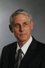
Speaker

Chair

Eli E Machtei

Harvard University, USA

Speaker

Co-Chair

Anderson de Oliveira Paulo

Faculty of Sciences of Tocantins (FACIT), Brazil

Speaker
Biography:

Eli E Machtei has obtained his DMD from the Hebrew University in Jerusalem and his certificate in Periodontology from Boston University. He is currently the Chairman of the department of Periodontology at the Rambam SGD and a Professor of Dental Medicine at the faculty of Medicine at the Technion (IIT) in Haifa Israel. He is also a Visiting Senior fellow at Harvard University. He has published over 170 papers in the scientific peer review literature and won several research awards for his work.

Abstract:

Implant placement marks the start of a chain of events in both the hard and soft tissues adjacent to the fixture. While earlier concepts have associated early bone loss around dental implants with physiological remodeling, more recent data suggests do not seem to support this notion anymore. Instead, early marginal bone loss is considered pathological and should be avoided or minimized. A multitude of factors have been suggested to be related to the prevalence and severity of early marginal bone loss around implants. These include placement protocol (submerged or non-submerged; crestal/supra or sub-crestal); loading protocols, implant's design and position, residual ridge's dimensions and the quality, width of the facial/lingual walls and the width and quality of the periimplant mucosa. Also, occlusal overload, crown-implant ration and implant's angulation, implants' diameter, flap versus flapless, mandibular versus maxillary implants and fixed versus removable implant supported restorations. Other systemic and environmental factors (smoking habits, diabetes, radiation therapy and past periodontal disease) were also suggested as possible risk factors. This presentation will focus on current knowledge regarding these variables and will try to highlight the currently available data along with many missing pieces in the puzzle. Finally, the ways to minimize this phenomenon will be discussed.

Speaker
Biography:

Anderson de Oliveira Paulo has completed his PhD at the Paulista State University (UNESP). He is Endodontics Professor of the Faculty of Sciences of Tocantins (FACIT). He is a Visiting Professor at the University of Brasilia (UNB). He has published several papers in journals. He is Co-author of a book and Author of five chapters in five books. In 2016, he was the President of the 8th International Congress of the Brazilian Society of Endodontics (SBENDO). He delivers lectures at congresses in Brazil and
in other countries.

Abstract:

For a long time, the endodontics was stagnant and without great prospects, it was a specialty seen as tiring for the professional and somewhat uncomfortable and even frightening for patients. All this began to change with the advent of NITI systems (Reciprocating and Rotary), with them, the root canal instrumentation became faster and stimulated the emergence of new root canals obturations techniques, These changes brought about a gain of time, a standardization of results. The advances were not limited to the root canal instrumentation and the obturation, they also brought benefits in the treatment of dental perforations, parendodontic surgeries and apical plugs with MTA. There were also advances in magnification with the lenses and especially with the microscope that allow the placement of channels, the visualization of separate instruments and perforations. Facilitating the treatment of each of these situations with the aid of ultrasonic tips of the last generation. And finally the appearance of the cone beam tomography that brought endodontics the possibility of seeing beyond the radiographic image. Because of all this, a better working condition for the endodontist, who ceased to be a sculptor and works with effective tools that did not require so much time of training and psychomotor improvement and the patients have access to faster and better-performing treatments with more comfort.

Speaker
Biography:

A Nayeemullah khan has completed his Under-graduation and Post-graduation from Meenakshi Ammal Dental College, Chennai. He is presently working as an Assistant-Professor in department of Orthodontics. He has published various articles in orthodontic journals. He is a vivid, resourceful, creative, solution oriented member of the team. The confidence which he shows in facing the varieties of challenges put forth against him, had brought praises from the members of the Cleft and Craniofacial Orthodontic Team. An enthusiastic clinician, he is always looking for higher challenges and stronger competitions, both nationally and internationally. He works effectively as a team member under the direction of other team leaders. His career in the institution and work in the surgical team truly reflect his academic standing.

Abstract:

Orthognathic surgery is gaining more positive response in India over the recent years owing to the increase in awareness to facial esthetics as the priority in undergoing treatment for skeletal malocclusions. Worsening of facial appearance during the period of pre-surgical orthodontics has been the reason for shift towards a surgery, first approach in the management of dentofacial deformities. A universal method for the use of surgery, first process is difficult to adapt as most patients worldwide treated by “surgery first” approach belong to skeletal Class III malocclusion. On the contrary, the Indian population showed diversity in ethnic facial pattern and presented more skeletal Class I and Class II malocclusions. The objective of this paper is to highlight  ifferences in treatment planning and execution of surgical first approach in severe skeletal Class I, Class II and Class III malocclusions and compare them to the conventional orthodontics first approach through ideal clinical scenarios and critically evaluate the early results achieved.

Mike Elvis Calderon

Calderon Institute, USA

Title: Demystifying the damon system
Speaker
Biography:

“After receiving his DDS Degree from the Dental School of Case Western Reserve Ohio in 1994, He moved back to Brooklyn, New York where he worked at a General Practice Residency at Kings County Hospital Between 1994 to 1999. Also Completing a 2 year program at the United States Dental Institute, where he was an orthodontics resident. Dr. Mike E. Calderón also completed 3 years at New York University as an Implant Resident. He has an extensive education and experience in Implant Dentistry (having placed more than 8000 implants). Dr. Calderón is a member of the Academy of General Dentistry, Diplomate of the ICOI, a Fellow and Diplomate of the American Academy of Implant Dentistry and Diplomate of the ABOI/ID. He established his private practice in Bay Shore, New York where he provides both advanced surgical and prosthetic phases of Implant dentistry for his patients. His ambition, sincerity, and passion for Dentistry are quite apparent when you meet him.

Abstract:

Introduction: The PRF (Platelet Rich Fibrin), PET (Partial extraction technique) also known as socket shield technique and incorporating everything the CTO Technique (Coronal Trans-Odontal Technique). These are three different procedures in the literature and refined to use together for better bone preservation. With all allogeneic materials other than the implant reducing cost and chance of rejection. Use of biological growth factors and ideal placement of the implant preparing the anatomical tooth before using the same as a guide for immediate placement all proves to be promising in osseointegration and bone preservation. All procedures were performed between two privately owned offices by the same surgeon. Methods: Pre and Post CBCT used for comparison. Only patients with conditions which contraindicated the placement of implants and without medical clearance were not treated. Results: Total of 39 implants, 30 Maxillary, 7 mandibular, 2 deciduous performed. 1 failure (97.4% success) after 2 years in function. Conclusion: Use of PRF was a breakthrough in implant dentistry. Socket shield technique proves to be another positive influence in Implant dentistry by helping prevent bone loss. The CTO Technique performed by two private clinics in NY proves to help properly place and implant the same day without the use of static guides. Use all three procedures prove to enhance osseoingration and proper placement of an implant in 3 dimensions.

Pankaj Ghalaut

Pandit Bhagwat Dayal Sharma Post Graduate Institute of Medical Sciences, India

Title: An Alternative to Conventional Dental Implants: Basal Implants
Speaker
Biography:

Dr. Pankaj Ghalaut currently serves as an Associate Professor at PGIDS, Rohtak , India. He has numerous publications to his name.

Abstract:

The conventional crestal implants are indicated in situations when an adequate vertical bone supply is given. These crestal implants function well in patients who provide adequate bone when treatment starts, but prognosis is not good as soon as augmentations become part of the treatment plan. Augmentation procedures tend to increase the risks and costs of dental implant treatment as well as the number of necessary operations. Patients who have severely atrophied jaw bones paradoxically receive little or no treatment, as long as crestal implants are considered the device of first choice. Basal implants are used to support single and multiple unit restorations in the upper and lower jaws. They can be placed in the extraction sockets and also in healed bone. Their structural characteristics allow placement in bone that is deficient in height and width. Basal implants are the devices of first choice, whenever (unpredictable) augmentations are part of an alternative treatment plan. The technique of basal Implantology solves all problems connected with conventional (crestal) Implantology. It is a customer oriented therapy, which meets the demands of the patients ideally. In this presentation the indications of using basal implants and the differences that exist between basal implants and crestal implants are discussed along with a case report.

Biography:

Zarina Paiziyeva has completed her DDS in the United States of America. Since 2012, she teaches at the Department of Dentistry at the Medical University of Astana. She has Graduated from the Masters in 2016, currently studying PhD in Doctoral studies. She studies the effective-ness of using a polysaccharide film in the  omplex treatment of oral dermatoses. For the period of scientific work, she has participated in  nternational conferences in London, Singapore, Dubai, Kazakhstan and  ublished more than 15 articles in journals

Abstract:

According to most authors, red flat lichen (RFL) is observed in many countries. Its prevalence ranges from 0.1% to 20%. The
incidence rate in different climatic geographic regions has signif-icant variations: from 45% in France, up to 0.13%-0.34% in
Sweden; In India, it is 1.4%. In the Republic of Kazakhstan, the frequency of the disease is 450: 100,000. The main place in the frequency
of localization of RFL is skin. Observations conducted indicate that of the 374 patients with RFL, there were manifestations of
oral mucosa in 54% of patients, while in 26.5% it was isolated. More recent studies show a lower incidence of RFL lesions in the oral mucosa: out of 585 patients, 22% of patients had eruptions on the oral mucosa, only 13.5% had an isolated le-sion of the oral mucosa alone. Red lichen is more common in people aged 30 to 50 years, in women more often than in men. Men get ill somewhat earlier (30 years) than women (40 years). The same researchers showed a tendency to "rejuvenate" this disease, which is probably due to the growing unfortunate environmental situation and the pace of life, coupled with a constant  sychoemotional stress. We have tested the effectiveness of the use of a   olysaccharide film and apparatus for phototherapy in the complex treatment of RFL. The effectiveness of treatment was evaluated in the immediate and long-term (12 and 24 months after treatment) for the dynamics of the pain syndrome,
the timing of epithelialization of lesions, the average duration of treatment,  hanges in cytological and immunological parameters, the frequency of relapse or exacerbation of the disease.

Biography:

Shady A. Moussa had received his Doctor of Dental Surgery in 2000 from Cairo University (Egypt) and his postgraduate Master of pediatric dentistry and oral public health from Al-Azhar University in 2006 and his doctorate DDS in pediatric dentistry and oral public health Al-Azhar University (Egypt) in 2012 also he earned MRACDS DPH (Australia) in 2017. He is a lecturer in Zagazig University, and consultant of pediatric dentistry in King Saud Hospital and King Fahad Specialist Hospital (Saudi Arabia). He is currently working as consultant of pediatric dentistry in (Qatar). Dr. Shady published several articles in peer-reviewed journals. He published a number of research papers in National and International Journals. He is an author of a one book. He is also an External Reviewer for a number of journals. Dr. Shady is an invited speaker to a number of national and international conferences and scientific meetings. He has presented seminars, lectures, continuing education programs and courses in pediatric dentistry and dental public health.

Abstract:

Background: Using video eyeglasses as an audiovisual distraction is useful in managing anxiety and reducing fear and anxiety in
healthy children during dental treatments. Dental anxiety has a significant impact on cooperative behavior during the dental visit,
particularly in children, and it could impede, or even preclude, the dental treatment. For these reasons, a knowledge of the appropriate
guidelines of behavioral management of children plays a key role in the oral health promotion and represents a major topic in
pediatric dentistry. Distraction is the technique of diverting the patient's attention from an unpleasant procedure.
Aim: The aim of this study is to evaluate the effect of audiovisual distraction on behavior and self-reported pain of children during
dental restorations and its influence on the operator stress and the time of the appointment.
Material & Methods: This randomized controlled crossover trial study of 96 outpatient’s healthy children under 12 years requiring at
least two dental fillings. The written informed consent for participation and publication was obtained from parents/legal guardians of each patient in full accordance with the ethical principles of the Helsinki Declaration. First dental filling was done wearing the videoeyeglasses and other one using conventional behavior management techniques. Subjective and objective pain was evaluated using the Faces Pain Scale - Revised (FPS-R) and the revised Face, Leg, Activity, Cry, and Consol ability scale (r-FLACC). The operator stress using a VAS, the time of the appointment, and the child satisfaction was recorded and tested by paired t-test. Results: Using video eyeglasses, significantly reduced the operator stress. The bivariate analysis showed that the mean FPS-R score and the mean r-FLACC score were significantly lower using the video eyeglasses, only during the second clinical session. Conclusion: Audiovisual distraction could be useful in managing anxiety in children but cannot replace the conventional behavior management techniques.

Speaker
Biography:

Sondus Al-Kadri, Syrian dentist has a Bachelor Degree in Dental Medicine from International University of Science and Technology, Damascus 2011. Practiced General Dentistry for two years in Saudi Arabia 2012-2013. Certified as a Clinical Orthodontic Assistant from International Training Institute (ITI), and worked as an orthodontic assistant in one of the prestigious Orthodontic clinics in “Kharsa Orhto-dent clinics” 2014. Got a full Master scholarship from Global Platform for Syrian Student, Portugal from 2015 - until now. Trained in Orthodontics Department at Malo clinics in Lisbon, 2015. Has a Master Degree in Dental Medicine from Faculty of Dental Medicine of Porto University, 2016. Now, she is pursuing her Master Degree in Orthodontics in University Institute of Health Sciences, (CESPU), Portugal.

Abstract:

Introduction: Bone-anchored maxillary expanders have been invented in order to provide extreme Pure skeletal expansion. There are studies that address the clinical effectiveness and outcomes of bone-borne maxillary expansion, but they were done mostly on adult subjects with surgical assistance. Objective: The main aim of this paper is to review publications that studied the effectiveness of bone-borne maxillary expansion compared with tooth-borne maxillary expansion in mixed dentition subjects. Search Strategy: A literature review using relevant articles dated from 2006 to June 2016 on PubMed and B-on databases was conducted. Relevant data were extracted, grouped and analyzed concerning three areas of research: the skeletal vs. dental expansion, side effects, and long-term stability of Tooth-borne maxillary expander (TBME), and Bone-borne maxillary expander (BBME). 16 articles were selected: 11studies addressed TBME; one study was a comparison between TBME and BBME, while the remaining 4 studies addressed BBME. Discussion: The outcome of the literature review showed that the percentage of skeletal expansion and dental expansion varied between TBME and BBME. Moreover, it also varied within TBME itself in different studies. Inter-coronal widths of maxillary permanent molars and first premolars in TBME were significantly greater than their counterparts in BBME. Generally, TBME showed a reduction in alveolar bone thickness on the buccal aspect of the banded teeth. Buccal tipping measured in TBME studies was small or high reaching up to 19 degrees, whereas in BBME group it was clinically trivial. Six months retention period is essential for recovery and long-term stability. Conclusion: Current available reviewed studies and clinical trials led to different inconsistent conclusions regarding the claim of the superiority and effectiveness of bone-borne versus tooth- borne maxillary expansion on mixed dentition patients. They were mostly done on adolescents or adults with surgical assistance. Although theoretically and logically this claim may seem valid, no evidencebased conclusion could be made since they were not done on patients below the age of twelve. Thus, future investigations on mixed dentition patients are highly recommended.

  • Prosthodontics | Dental Implantology | Laser Therapy in Dentistry | Dental Ethics & Public Health | Dental Marketing & Management | Oral & Maxillofacial Surgery
Location: Tribeca II
Speaker

Chair

Peter Bongard

MVZ Zahn+Zentrum Moers, Germany

Speaker

Co-Chair

Nayeemullah Khan

MAHER University, India

Session Introduction

Peter Bongard

MVZ Zahn+Zentrum Moers, Germany

Title: Implants in the aesthetic zone. How to make the right decision?
Speaker
Biography:

Peter Bongard is a renowned speaker in the field of laser dentistry. He completed his higher education entrance certificate from Gymnasium Adolfinum, Moers in 1988. He also underwent apprenticeship as a paramedic during 1986-1988. He obtained his bachelor’s degree in dentistry in 1993 from Westfälische Wilhelms Universität ,Münster. He has served as an Assistant Doctor at Joint practice Dres. Klotz / Zückler, Duisburg between 1993 – 1995. His interests include periodontology & Laser dentistry.

Abstract:

Success is the summation of missteps that have not been made. (Albert Einstein). Before starting the therapy, it is most important to analyses the correct time for the extraction, the implantation, the augmentation and the connective tissue graft up to the decision for the ideal abutment and the prosthetics. The key point for all this is a special aesthetic analysis at the very beginning. The lecturer presents a master plan for the perfect aesthetics in the high-risk zone and a strategy to avoid periimplantitis.

Speaker
Biography:

Liciane Toledo Bello has completed her Master Science in Dentristy. She is working in Dentistry Lasers since twenty years. She is a Clinical Director in the Dentistry departement of the Neomama Institute.

Abstract:

The Modified ILIB, is a modification of original therapy, which introduces a glass fiber into the blood vessel for emission laser radiation. In the modified technique the irradiation is transdermical bringing benefits in realtion to the inflammatory marker. In this case report, over three years, patient MSL, woman with eighty-one years, received ILIB, demonstrated seric lebes of the reactive C protein, starting ar approximately 13 mg/l and gaving this index reced to1,38 mg/l two years later, without any cardiovascular events.

Aye Myat Thwe

University of Dundee, United Kingdom

Title: Role of EGFR inhibitors in oral cancer cell migration
Biography:

Aye Thwe graduated from Myanmar with a Bachelor of Dental Surgery in 2010. After practicing as a Dentist for 2 years, she came to UK to study at University of Dundee. She received an MRes in Oral Cancer, and progressed into the PhD programme. She is now in the 3rd year of her PhD programme.

Abstract:

Epithelial to mesenchymal transition (EMT) is the process by which cells change shape from being tightly connected epithelial cells to more motile mesenchymal cell. EMT has been reported to facilitate cell migration. Cell motility is an initial first step on the road to metastasis. Epidermal growth factor receptor (EGFR) has been reported to be overexpressed in oral cancer and is often related with poor prognosis. Epidermal growth factor (EGF) and transforming growth factor (TGFα) are ligands that bind to EGFR and can affect many different cellular processes such as proliferation, migration, apoptosis etc. In this project, cell proliferation, migration, morphology change and EMT makers of HSG, AZA1, HacaT and TYS are measured by cell counting, scratch assay, photographic image capturing and  mmunofluorescence in related with addition of 1 ng/ml, 10 ng/ml, 50 ng/ml of EGF and TGFα incubated at different time point. 10 ng/ml and 50 ng/ml concentration induce morphology change (EMT like phenotype with finger like projection) and increase migration while there is not much difference in cell proliferation. Their morphological changes are completely blocked by 1hour pre-treatment with 5μM Gefitinib (EGFR tyrosine kinase inhibitor), 5 μM Erlotinib (EGFR TK inhibitor) and 25 μM PD (MAPK inhibitor) while there is no blockage of cell migration.

Biography:

Jessica Sidharta was the head of an esthetic dental surgery in Munich with a focus on veneers and bleaching. She is a Lecturer and KOL of edelweiss dentistry. She used edelweiss VENEERs on a regular basis in her own dental office. Nowadays she uses the VENEERs regularly at the edelweiss dentistry in Wolfurt/Austria. Jessica Sidharta had further education with Prof. Dr. Dr. Didier Dietschi and since 2012 she is an established dentist in Ulm with a focus on esthetics.

Abstract:

After different experiences with ceramic restorations, I made amazing experiences with the edelweiss direct veneer system. This system makes your every-day-dentist-work much more easier. Giving your patients a natural bright smile is a fulfilling satisfaction for you as a dentist and of course for your patient. For every situation I have all solutions in my edelweiss direct system every time. With the possibility of every colour individualization and every morphology I can give the patient a beautiful smile back in one appointment, which safe time and costs for the patient and for the dentist.” Jessica Sidharta D.M.D.C.D.T “The edelweiss direct system works together with the teeth, the result is one monoblock in form and function.”
EVERY DAY EXPERIENCE WITH EDELWEISS DIRECT SYSTEM
My experience in practice was that: Often at the end of the day or of the week, you have patients after accidents, mainly young people
with broken anteriors after bike accident or jumping into a swimming pool. With the edelweiss system I have the possibility to give
them smile back in just one appointment without exaggerated costs. Because of the material, which is laser-sintered composite, you can treat minimally-invasive or even non-invasive. The material is not ceramic, that is why you do not need minimal thickness. Necessary grinding can be done on the veneer directly. The material is biocompatible and has the same mechanical features like our teeth. So the dentists have also a solution for patients suffering of bruxism, because of the same flexural modulus like the natural teeth. This system works together with the nature, it is not too much stiff like ceramic. All grinding individualizations and morphology designs are possible. After short polishing you get the perfect shining back. All products of edelweiss dentistry never changed the colour in my experiences since 2011.

Biography:

Dr. Mashael Bin Hasan graduated with a bachelor’s degree in dentistry from King Saud University in 2004. She completed her masters in operative dentistry in 2010 from the King Saud University where she currently serves as a fulltime lecturer on operative dentistry.

Abstract:

The last two decades have evidenced a paradigm shift in the dental practice that has been guided by a greater understanding of science. There has been a change from patients seeking treatment to elective cosmetic enhancement. The concern in health and beauty has become an important factor that increased the demand for cosmetic dental procedures. In the past, extensive, invasive and expensive dental procedures were required to achieve a beautiful smile. Nowadays, the evolution in restorative materials, adhesives and technologies expanded the treatment  ossibilities for the clinician, treatment options available for the patients and provided simple, conservative and economical solutions to many of the restorative and esthetic challenges.

  • Periodontology | Dental Ethics & Public Health | Fundamental Dentistry | Oral & Maxillofacial Surgery
Location: Tribeca-II

Session Introduction

Harvinder Singh

Maharishi Markandeshwar University, India

Title: Balanced Smile: A perspective for orthodontic treatment planning
Biography:

Dr. Harvinder singh has completed his BDS at the age of 26 yrs from Maharishi Markandeshwar University, Mullana, India. At present he is a final year Post Graduate student in the Department. of Orthodontics and Dentofacial Orthopedics, Maharishi Markandeshwar University, Mullana, India. He has presented a clinical poster and a scientific paper at IOS PG Convention at Amritsar and Banaras respectively and one poster at annual conference of Indian Society of Cleft Lip Palate & Craniofacial Anomalies.

Abstract:

Facial esthetics has been the most important objective of orthodontic treatment planning. Smile characteristics are determined
by static and dynamic relationships between hard and soft tissue components of face. The objective of the present study was to
evaluate the balanced smile using different hard and soft tissue parameters.A total of 50 participants equally divided into two goups i.e
males and females, ranging in age from 18–25 years with Angle’s Class 1 molar relation were selected. Photographic records of smile
and rest position were measured and analyzed at rest and during smile, to assess upper lip length, maxillary incisal display, upper
vermillion length, intercommissural width and smile arc. Results obtained from both the groups were subjected to stastical analysis
and correlations between smile variables were investigated. Most of the measured variables showed statistically significant sexual dimorphism. Statistically significant results were seen in relaxed upper lip length, relaxed central incisor display, relaxed vermillion length, smiling upper lip length and smiling vermillion length. Resting inter commissural width, smiling central incisor display and smiling inter commissural width were found to be statistically non-significant. Upper lip length and vermillion length in smiling and resting conditions had same pattern. Increased incisal display was more evident in subjects with short upper lip. Parallel smile arc and flat smile arc distribution was more common in females subjects whereas reverse smile arc was seen in male patients only.

Biography:

Diaa E Mostafa is an Assistant Lecturer in the oral and maxillofacial surgery department of faculty of Dentistry at the University of Minia, Egypt, where he has been a Faculty Member since 2007. He has completed his MSc and  undergraduate studies at Minia University. His research interests lie in the area of temporomandibular joint disorders. In recent years and for his PhD research, he has focused on better techniques for arthroscopic management of TMJ. He has served on many conference and workshop program committees. He is an Instructor for a clinical part of oral surgery for undergraduate’s topics in his department and Vice Director of cranio- maxillofacial surgery unit at Minia University Dental Hospital.

Abstract:

Mouhibi Abdallah

Casablnca University of Hassan II, Morocco

Title: The place of peek in evidence based dentistry and implantology
Biography:

Mouhibi Abdallah has completed his Dental medicine at the age of 22 years from Faculty od Dental Medecin of Casablnca from Hassan II University of  Casablanca, and pursuing residant in Prosthodontics.

Abstract:

The PEEK Medical Polyetheretherketone is a high performance polymer. The materials in this group combine excellent mechanical properties with the highest biological compatibility. PEEK is also used in aeronautical construction for heavy parts and for medical applications. It has evolved in the field, especially for the manufacture of denture frames. The elasticity of the material, which is of the same order as that of bone tissue, makes it a nature-like material since it can compensate for bone torsion, especially in the presence of large implantation work. It has replaced titanium with its properties and qualities. This work aims to demystify this material by demonstrating its advantages and limitations and its place in current dentistry.

Malak Nagi

Malak Nagi Dental Clinic, Egypt

Title: Frame it
Biography:

Malak Nagi has completed his BSc in Dental Medicine and Oral Surgery in 1995 from Alexandria University and DDS in Pedodontics in 1998 from Alexandria University, Egypt. He is the owner of Malak Nagi Dental Clinic in Alexandria. He is a board member of Alexandria Oral Implantology Association and International College of Dentists “Region 29”. He is a Lecturer in Facial Esthetics “Botox & Fillers”.

Abstract:

As a dentist you must keep in mind that you are creating a piece of art with each case you are treating. Frame it is a course that gives you the fundamental keys for creating a beautiful frame for your patients’ teeth using Botulinum Toxin A and dermal fillers. The course will cover several topics including; facial anatomy for various cosmetic applications, Botulinum Toxin A (mechanism of action, proper storage, preparation, indications, contraindications, precautions for use, proper management of complications and injection techniques), dermal fillers (characteristics of various fillers available in the market and pain management options), Hyaluronic acid filling agents (indications, contraindications, precautions for use, proper management of complications and injection techniques), as well as, the role of Golden Ratio in facial esthetics. By the end of the course, the participant should be able to manage basic cases that need a fine touch to reach optimal esthetic results.

Biography:

Dr.Puneeta Vohra Associate professor in SGT University Gurgaon India since 2014. Received BDS degree form Himachal University India 2007 and masters from Sumandeep Vidyapeeth university Gujrat in 2010, India. Her main interest is evaluation of patients with HIV and has to her credit publications as well paper presentation at national level. Presently she is associated with teaching undergraduate levels as well as guiding post graduates in Oral Medicine and Radiology stream”.

Abstract:

HIV infection is a major health concern in India. India has the third highest number of people living with HIV in the world with 2.1 million Indians accounting for about four out of 10 people infected with the deadly virus in the Asia Pacific region. There are considerable regional variations in the oral manifestations of HIV infection, depending both on the populations studied and on the clinical heterogeneity. The oral manifestations in Asian countries as compared to western countries and other developing nations in Africa and Latin Americas are relatively different. The constraint of resources in Asian countries hinders the possibility of providing an effective health care system. Though many studies have been conducted on oral manifestations in HIV, the research in Asia is currently at a pivotal juncture as the existing research is getting repetitive. Currently, Asian studies predominantly focus on prevalence of oral manifestations and assess their association with parameters of HIV infection, sociodemographic data and deleterious habits. Hence a cross sectional study was undertaken among HIV population in western India, Gujarat with main focus and objectives: 1) Evaluation and diagnostic usefulness of saliva for detection of HIV antibody 2) To calculate CD4 count of HIV positive patients. 2) To evaluate systemic manifestations of HIV positive patients. 3) To detect oro-maxillofacial manifestations in HIV positive patients. 4)To compare and correlate systemic and oro-maxillofacial manifestations of HIV positive patients with the CD4 count. Study population comprised of newly diagnosed 100 confirmed seropostive patients, before starting any antiretroviral therapy, were included. Saliva of HIV positive patients was collected and was tested by ELISA by using special reagents and modifications leading to detection of antibody in saliva. The oral lesions were diagnosed based on clinical manifestation using international criteria and CD4 count was determined within maximum 1 week of oral examination. Oral and systemic manifestations of HIV positive patients were recorded and correlated with CD4 counts. It was found that decrease in CD4 count is associated with a wide range of oral and systemic manifestations which can be used as prognostic marker for immune suppression in AIDS patient. This study can be very useful in epidemiological surveys, diagnosis and management of HIV positive patients.